
Contents (Jump to a section)
Can Tight Calves Cause Knee Pain? The Calf-Knee Link Explained
Introduction
Yes, tight calves can cause knee pain. This is not a vague correlation. There is a direct mechanical explanation: the gastrocnemius, the large superficial calf muscle, originates from the posterior femoral condyles, directly above the knee joint. When it is chronically shortened, it pulls on the back of the femur and creates compressive forces at the knee. At the same time, restricted ankle dorsiflexion from calf tightness forces compensatory movement patterns that load the knee in mechanically disadvantaged positions.
Most people experiencing anterior knee pain, patellar irritation, or diffuse knee aching are not told to check their calf flexibility. The focus goes straight to the knee itself: quad strength, patellar tracking taping, or imaging. But if the calf is the source of altered mechanics, treating the knee without addressing the calf misses the root cause.
This article explains the calf-knee biomechanical chain from the ground up, identifies the specific movement patterns that connect calf tightness to knee pain, and provides practical stretching protocols to address both muscles involved.
The calf-knee anatomy you need to know
The calf complex consists of two anatomically distinct muscles: the gastrocnemius and the soleus. Understanding what each one does, and where each one attaches, is the foundation for understanding why calf tightness affects the knee.
The gastrocnemius is the larger, superficial muscle that creates the characteristic bulge at the back of the lower leg. It originates from the posterior femoral condyles, just above the knee, and inserts via the Achilles tendon into the heel bone (calcaneus). Because it crosses both the ankle and the knee joint, it is a two-joint muscle. This is the critical anatomical fact: when the gastrocnemius is shortened, it does not just restrict the ankle. It also pulls on the femoral condyles, applying a posteriorly directed force at the knee.
The soleus lies beneath the gastrocnemius and originates from the posterior tibia and fibula, below the knee. It also inserts via the Achilles tendon. The soleus does not cross the knee, so it does not directly pull on the femoral structures. However, it is a powerful restrictor of ankle dorsiflexion, the upward motion of the foot toward the shin. Limited dorsiflexion from soleus tightness forces the knee to compensate during every step.
Both muscles share the Achilles tendon, and chronic tightness in either, or both, creates mechanical consequences that travel upward through the tibia and into the knee joint. Understanding which muscle is tighter tells you which stretch you need.
How tight calves alter knee tracking
Knee tracking refers to the path the patella follows as the knee flexes and extends. Under normal mechanics, the patella glides in a vertical groove on the femur. When calf tightness restricts ankle dorsiflexion, the body finds compensatory pathways to absorb load, and those pathways stress the knee.
The most common compensation is excessive pronation at the foot and ankle. When you cannot dorsiflex adequately, the foot collapses inward to gain the range of motion the ankle cannot provide. This inward rolling of the foot externally rotates the tibia, which in turn causes the knee to track medially rather than straight. The patella now moves in a path it was not designed for, creating irritation at the lateral patellar facet and in the soft tissue structures that guide patellar movement.
A second compensation involves forward lean of the trunk and increased knee flexion under load. Squatting, stair climbing, and descending a slope all require adequate ankle dorsiflexion. When that range is absent, the tibia cannot travel forward over the foot, so the knee absorbs extra shear force and the quadriceps tendon works at a less favorable angle. Over time, this repetitive loading pattern contributes to patellar tendinopathy, patellofemoral pain syndrome, and generalized anterior knee aching.
A third mechanism is the direct compressive effect of the gastrocnemius. During movements that load the knee in a bent position, a tight gastrocnemius increases the posterior force on the femoral condyles. This effectively increases the compressive load within the joint at precisely the angles where tissue damage accumulates.
Specific conditions linked to tight calf muscles
Tight calves have a documented association with several common knee complaints. Knowing which condition connects to which mechanism helps you target your approach.
Patellofemoral pain syndrome
Patellofemoral pain syndrome, often called runner's knee, is characterized by aching around or behind the kneecap, particularly during stair climbing, squatting, or prolonged sitting. Restricted ankle dorsiflexion from calf tightness is one of the contributing mechanical factors. When the tibia cannot travel forward adequately during a squat or step, the knee compensates by allowing the patella to track laterally. Improving dorsiflexion through targeted calf stretching reduces this compensatory pattern.
Patellar tendinopathy
Patellar tendinopathy involves degeneration and pain of the tendon connecting the kneecap to the shinbone. Restricted dorsiflexion forces the knee into positions of higher tendon stress during landing, jumping, and deceleration. Athletes with tight calves who jump repeatedly, basketball and volleyball players in particular, have elevated tendinopathy risk because each landing compresses the tendon at a suboptimal angle.
Iliotibial band syndrome
Iliotibial band syndrome causes lateral knee pain during running. Tibial rotation from calf-driven overpronation is one of the upstream contributors. When the foot collapses inward because ankle dorsiflexion is restricted, the tibia externally rotates, increasing the tension and friction at the point where the IT band contacts the lateral femoral condyle. Addressing calf tightness as part of an IT band protocol, rather than focusing on the IT band alone, often produces better and faster results.
Generalized knee aching in desk workers
People who sit for long periods often develop diffuse knee stiffness, particularly noticeable when standing after extended sitting. Prolonged sitting keeps the knee flexed and the ankle plantarflexed, which shortens the calf complex in both positions simultaneously. When that person then stands and walks, restricted dorsiflexion alters the gait cycle and increases loading at the knee. This pattern is frequently misattributed to aging or arthritis when calf tightness is actually the primary driver.
The dorsiflexion restriction test
Before you begin stretching, it is worth assessing where your ankle dorsiflexion actually stands. A simple self-assessment: stand facing a wall, place your big toe 10 centimeters (about 4 inches) from the wall, and try to touch your knee to the wall without lifting your heel. If you cannot touch the wall with your knee at that distance, you have restricted dorsiflexion, and your calf tightness is likely influencing your knee mechanics.
Most adults with adequate dorsiflexion can pass this test at 10 to 12 centimeters. Athletes in high-demand sports should ideally pass at 12 to 15 centimeters. The test is not a diagnosis, but it gives you a concrete baseline to track as your calf flexibility improves. Many people are surprised to discover that significant restriction exists on one side but not the other, and that this asymmetry corresponds directly with the side where they experience knee complaints.
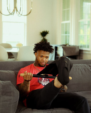
💠 For people managing chronic calf tightness linked to knee discomfort, heat combined with targeted stretching can accelerate tissue response. The CastleFlexx Heat Edition is the only heated stretch device in the world, applying consistent therapeutic warmth to the ankle and calf complex while guiding the stretch into the correct dorsiflexion angle. It is a patented, USPTO-registered device used in home recovery programs to help treat the root cause of lower-leg tightness and the knee mechanics it affects. Learn more at castleflexx.com.
Targeted stretching protocol for the calf-knee chain
Addressing calf tightness in the context of knee pain requires targeting both the gastrocnemius and the soleus. Each needs a different technique because the gastrocnemius crosses the knee and the soleus does not.
Wall calf stretch (gastrocnemius)
Face a wall and place both hands at shoulder height. Step one foot back approximately 2 to 3 feet, keeping the heel firmly planted and the back knee straight. Lean your hips toward the wall until you feel a clear stretch running from the heel through the calf belly. Hold for 45 seconds per side, performing 3 sets. The heel must stay on the ground at all times. If it rises, the stretch is lost.
The straight back knee is essential for isolating the gastrocnemius. This is the muscle that crosses the knee and directly affects knee mechanics. Prioritize this variation if your knee symptoms are related to patellar tracking or patellofemoral pain.
Bent-knee calf stretch (soleus)
Use the same wall position as above but bend the back knee 15 to 20 degrees rather than keeping it straight. This slackens the gastrocnemius and shifts the load to the soleus. You will notice the stretch sensation moves from the upper calf toward the lower calf and the area just above the heel.
Hold for 45 seconds per side, 3 sets. People with Achilles tightness often find this variation produces a more direct therapeutic sensation. Include both variations in every session for complete coverage of the calf complex.
Stair calf stretch
Stand on a step holding the railing for balance. Position the ball of one foot on the step edge with the heel hanging off. Allow the heel to lower slowly toward the floor, controlling the descent. Hold for 30 to 45 seconds per side, 3 sets. This variation achieves a larger dorsiflexion range than flat-surface stretches and provides an excellent eccentric stimulus when you control the lowering phase.
Introduce this variation after 1 to 2 weeks of foundational wall stretching. In the early sessions, lower the heel only partially. Over time, work toward a full-range heel drop.
Downward dog
Start on hands and knees. Press your hands firmly into the floor, lift your hips upward and back, and form an inverted V shape. Work to press your heels toward the floor. Hold for 45 to 60 seconds, repeating 2 to 3 times. This variation stretches both calves simultaneously and allows you to alternate pressing one heel down while lifting the other, creating a dynamic stretch across the entire calf complex.
Frequency and progression for knee-related calf tightness
When calf tightness is contributing to knee pain, daily stretching is more important than intermittent sessions. The nervous system and connective tissue both respond to frequency. Stretching three times per week produces slower results than short daily sessions.
A practical protocol: perform the wall stretch (both straight and bent-knee versions) twice daily, morning and evening, for the first two weeks. Each session takes 6 to 8 minutes. After two weeks, add the stair stretch and downward dog. By week four, most people notice improved ankle range and reduced knee symptoms during daily activities.
Progress is tracked with the dorsiflexion wall test. Repeat the assessment every two weeks. Most people see the distance increase by 1 to 2 centimeters per testing period when stretching consistently. As dorsiflexion improves, knee mechanics during squatting and stair climbing typically improve as well.
Pair stretching with calf strengthening. Standing calf raises, both straight-knee and bent-knee versions, build the strength needed to support a more mobile calf through the full range of motion. Flexible but weak calves are prone to re-tightening under load. Strengthening locks in the gains from stretching.
Frequently Asked Questions
Can tight calves cause knee pain?
Yes. The gastrocnemius originates from the posterior femoral condyles, just above the knee. When this muscle is chronically shortened, it applies a posteriorly directed compressive force at the knee. Additionally, calf tightness restricts ankle dorsiflexion, which forces compensatory movement patterns during squatting, walking, and stair climbing that load the knee in mechanically disadvantaged positions.
What knee conditions are linked to calf tightness?
Patellofemoral pain syndrome (runner's knee), patellar tendinopathy, iliotibial band syndrome, and generalized anterior knee aching are all associated with restricted ankle dorsiflexion from calf tightness. Improving calf flexibility is a standard component of rehabilitation for these conditions.
How do I know if my calves are affecting my knee?
Perform the wall dorsiflexion test: place your big toe 10 centimeters from a wall and try to touch your knee to the wall without lifting your heel. If you cannot, your dorsiflexion is restricted and your calf tightness is likely altering knee mechanics. Asymmetry between sides often corresponds with the side where knee symptoms occur.
How long does it take for calf stretching to improve knee pain?
Most people notice improved ankle range within 5 to 7 days of consistent daily stretching. Changes in knee mechanics and symptom reduction typically follow within 3 to 4 weeks when stretching is combined with gait awareness and calf strengthening. Structural tissue changes take 6 to 8 weeks of consistent work.
Should I stretch both calves even if only one knee hurts?
Yes. Asymmetric flexibility is common, but both calves should be addressed. Stretching only the symptomatic side leaves the unaffected side vulnerable to developing tightness and alters pelvic and knee mechanics through gait compensation. Always stretch bilaterally.
Does calf tightness cause the same knee pain as a meniscus injury?
Not the same pain. Meniscus injuries typically produce localized joint-line pain, swelling, and mechanical catching or locking. Calf-related knee pain is usually diffuse, anterior, or around the kneecap, and worsens with repeated loading like stair climbing or running rather than with a specific incident. If you are unsure of the cause of your knee pain, see a physical therapist or orthopedic physician for assessment.
Key Takeaways
- The gastrocnemius originates above the knee, meaning calf tightness applies direct compressive force at the knee joint and is not limited to ankle restriction.
- Restricted ankle dorsiflexion from calf tightness forces compensatory pronation and tibial rotation, altering patellar tracking and increasing load on the knee's soft tissue structures.
- Patellofemoral pain syndrome, patellar tendinopathy, and IT band syndrome all have calf tightness as a contributing upstream factor.
- Target both muscles: straight-knee wall stretches for the gastrocnemius, bent-knee stretches for the soleus. Each requires a separate technique.
- Daily stretching, twice per day during the first two weeks, produces faster results than three sessions per week when knee symptoms are the goal.
- Pair calf stretching with calf strengthening to lock in flexibility gains and prevent re-tightening under load.
When to See a Professional
Calf stretching is appropriate for most people experiencing knee discomfort related to mechanics and tightness. Consult a physical therapist or orthopedic physician if knee pain is sharp, accompanied by swelling or warmth, follows a specific traumatic incident, includes joint-line tenderness, or does not improve after three to four weeks of consistent calf stretching and strengthening. These signs may indicate meniscal, ligamentous, or cartilage pathology that requires clinical assessment beyond a self-directed stretching program.
About the Reviewer
Dr. Sarah Patel, DPT, SCS, is a licensed Doctor of Physical Therapy with a board certification in Sports Clinical Specialty. She has over twelve years of clinical experience working with competitive runners, endurance athletes, and active adults recovering from lower-extremity and knee conditions. Dr. Patel completed her clinical residency in sports rehabilitation and reviews CastleFlexx content to ensure clinical accuracy and appropriate scope of guidance.